What is a Lung Nodule?
Lung nodule is a remnant of a lung infection smaller than 4 cm in diameter, a benign lung tumor, or lesions that occur with the spread of lung cancer or another organ cancer to the lung. Lung nodules are also popularly defined as “spots on the lung”.
Lung nodules encountered in radiological imaging results:
- Solitary nodule
- Semi-solid nodule, half-frosted glass nodule
- Frosted glass nodule
- Several nodules
The presence of nodules in the lung is evaluated considering the following conditions:
- Patient’s age
- Smoking history
- Previous diseases
- Have a relative with lung cancer
- Whether this nodule exists in his previous films
- Needle biopsy result
- PET-CT findings
As a result of these evaluations:
- A completely benign nodule
- Suspicious nodule
- Lung cancer suspected is classified as a very high nodule.
What is the Frequency of Pulmonary Nodules?
On average, 30% of smokers over the age of 55 have lung nodules.
Only 1.5% of these patients develop cancer in these nodules.
The important thing here is to detect this nodule, which has a 1.5% probability of being cancerous. All of our work and the website are to guide you in this regard.
The following 2 conditions are important: 1) The patient and his family have a suspicious life for a simple nodule in the lung. 2) Although the nodule in the lung is indeed a suspicious lesion for cancer, it is not taken seriously and it grows and spreads (metastasizes).
Malicious Lung Nodule
The probability of a lung nodule being malignant, meaning cancer:
- If the nodule is smaller than 5mm, the probability of it being a cancer is 0% – %1
- Nodule 5 – If between 10 mm: 6% – 28%
- If the nodule is larger than 2cm: 64% – 82%
- Probability of cancer in a solid-looking nodule: 7-9%
- Probability of cancer in a ground glass nodule: 59-73%.
(Source: Chest Journal. September 2007, Vol 132, No. 3_suppl)
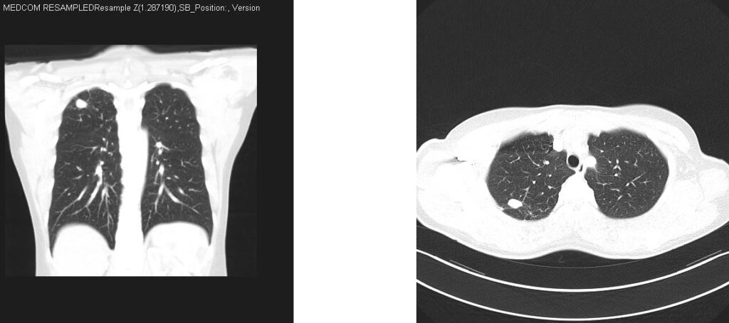
In the picture below, there is a computed tomography image of a 67-year-old female patient with a ground-glass nodule in the upper lobe of the right lung. As a result of the surgical removal of the nodule, it was determined that it was an adenocarcinoma and lobectomy was performed.
Benign Lung Nodule
The nodule in the lung can be decided as a benign nodule under the following conditions:
- Patient younger than 30 years
- Nodule smaller than 4mm
- Nodule in solid form, not in ground glass appearance
- Presence of a nodule of the same diameter in previous chest X-ray or tomography
- No high uptake in PET-CT images
- No relatives with lung cancer
- The patient has not had any other organ cancer before
- No cancer was found as a result of needle biopsy from the nodule
- The presence of calcification in the nodule
There is no treatment required for these nodules that are determined to be benign.
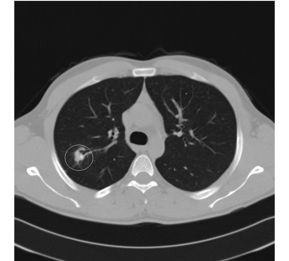
In the example below, a nodule located in the upper lobe of the right lung is seen in a 34-year-old female patient who has never smoked. With the evaluations and 3-month follow-ups, it was understood that this nodule was benign. Our patient has been under follow-up for more than 3 years and is in good health.
How to Identify a Malignant Nodule in the Lung?

Patient-related conditions
- Patient age greater than 40
- Being a current or former smoker
- Lung cancer in close relatives
- The patient has had another cancer before
Conditions about the nodule
- Nodule larger than 1cm
- Has a frosted glass look
- Irregular borders
- Absence of lung nodules in previous films
- The fact that the diameter of the nodule present in the previous films has grown
- Increased activity in the nodule in PET-CT (as seen in the picture above)
Which Lung Nodules Should Be Removed Surgically?
In cases with a high risk of Lung Cancer, the nodule should be removed endoscopically and examined by pathologists, and a definitive diagnosis should be made.
What do International Sources recommend?
Lung Nodule Surgery (Single Port VATS)
This procedure is performed endoscopically (closed surgery) with a method called VATS with a 2 cm incision. Removal of the nodule is performed with a camera and surgical instruments advanced into the chest cavity through this incision. The removed nodule is sent for pathology examination during the procedure and the result is lung cancer lobectomy is performed in the same session. If it is reported that there is no cancer in the nodule, there is no need for lobectomy.
How Many Days Should I Stay in the Hospital for Surgery?
After nodule surgery with VATS (Closed Surgery), patients are discharged within 1-3 days and return to work 3 days later. If it is detected that there is cancer in the nodule, then the lobectomy will be performed during the same operation, so the discharge is 3 to 5 days, and the return to work is 10 days later.
NEW: In June 2015, the British Thoracic Society published a highly detailed guide for the diagnosis, follow-up and treatment of pulmonary nodules. Most of the information you read on this website is based on the recommendations in this guide. To access this guide in English, You can visit http://thorax.bmj.com/content/70/Suppl_2/ii1.full.pdf+html.